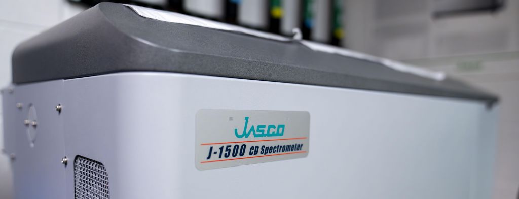
Summary Circular Dichroism (CD) is an absorption spectroscopy method based on the differential absorption of left and right circularly polarized light. CD is used to determine aspects of protein secondary and tertiary structure.

Uses
- Identify structural aspects of proteins and DNA
- Secondary structures of proteins can be analyzed using the far-UV (190-250 nm) region
- The ordered α-helices, β-sheets, β-turn, and random coil conformations determined in far-UV region
- Study conformational changes in molecules due to different buffer conditions
- Show the extent of denaturation with a change in temperature or chemical environment
- It can also provide information about structural changes upon ligand binding
- Information about the tertiary structure of proteins can be determined using near-UV spectroscopy (250-300 nm) of the dipole orientation and surrounding environment of the aromatic amino acids.
Specifications
- Xenon arc lamp source
- Double monochromator offers low stray light
- Nitrogen purge offers even far UV CD applications feasible
- Two quartz cell path lengths, 1mm and .1mm possible
- Temperature-wavelength scan measurement
- Temperature ramping (-40 to 130°C) and thermal denaturation for screening binding
- Wide spectral range from vacuum UV to Near-IR (163 to 1600 nm)
- Standard built-in mercury lamp and optional NIST traceable standard sample for system validation
- High-efficiency N2 purge capability enabling to enhanced inert UV measurement
- Extremely low stray light and high S/N ratio providing wide dynamic range
- High speed scanning (10000 nm/min)
- Spectra Manager II - Software Suite for data acquisition, analysis and presentation including several methods of secondary structure calculation
Details
Buffer Choice: Many common buffer components absorb strongly, particularly at shorter wavelengths, and can mask signals of interest. In general, try to keep buffer concentrations as low as possible and observe the following:
- 10mM potassium phosphate is a good choice for most work. Low concentrations of perchlorate, Tris, sodium phosphate and borate are also fairly transparent.
- Cl- has a strong UV absorbance at low wavelengths. SO42- or F- is a preferred counter ion.
- DTT, BME, or EDTA can be present at low concentrations (≤1mM).
- Imidazole absorbs strongly in the far UV. Even millimolar concentrations of imidazole will swamp your micromolar protein signal.
- Buffers can contain up to 20% glycerol, but measurements can only be made to 200 nm at this concentration.
- SDS, Chaps and octylglucoside are reasonably transparent detergents. Avoid Triton detergents as they tend to oxidize rapidly and form UV-absorbing materials.
- The pH of Tris is highly dependent on temperature, which makes it a poor choice for thermal melts.
- For denaturant melts, be sure to use ultrapure spectral grade urea or guanidine.
- Use UV=grade solvents.
- Avoid using water that has been stored in polyethylene bottles for a long time as it may contain dissolved polymer additives.
Filtration: Filtering your samples and buffer through a 0.2µ or 0.45µ filter is a good way to remove dust, aggregated protein and other particles that interfere with CD measurements.
Cuvette Path length:
- Smaller pathlength cuvette (1mm) decrease solvent absorbance and is used for far-UV measurements.
- A typical concentration for a 1 mm cell is ~0.1 mg/mL
- Minimum volume ~200µL
- Longer pathlength cuvette (10mm) is used for near-UV measurements since the signals are usually weak.
- Near-UV measurements typically require high concentrations, ~1-2 mg/mL
- Minimum volume of the standard cell is ~2mL
Approximate wavelength cutoffs (nm) of various solvents/buffers
| 1 mm cuvette | |
| Distilled Water | <185 |
| 100mM Ammonium Citrate | 220 |
| 150mM Ammonium Sulfate | 190 |
| 100mM Mes | 205 |
| 100mM Pipes | 215 |
| 100mM Sodium Chloride | 195 |
| 10mM Sodium Phosphate | <185 |
| 100mM Sodium Phosphate | 190 |
| PBS (phosphate buffered saline) | 200 |
| 100mM Tris-HCl | 200 |
| Acetonitrile | 185 |
| DMSO | 252 |
| Ethanol | 195 |
| Hexafluoroisopropanol | <185 |
| Methanol | 195 |
| Trifluoroethanol | <185 |
| 4M GdnHCl | 210 |
| 4M Urea | 210 |
Acknowledgement
Please acknowledge us as “Penn State X-Ray Crystallography Facility - University Park"