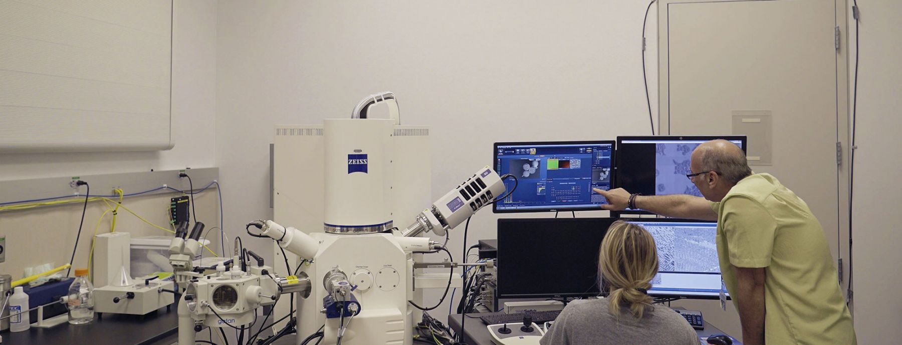
The Microscopy Facility specializes in three areas: optical microscopy, electron microscopy, and histology. The facility is equipped with confocal microscopes, research fluorescence microscopes, and transmission- and scanning-electron microscopes. Its research staff engage in experimentation, training, project collaboration, and consultation.

Microscopy Facility
Employing optical and electron microscopy and histology instrumentation, along with expert assistance, to advance your research
News
Microscopy director propagates new discoveries and new scientists
The directors of the core facilities at Penn State’s Huck Institutes of the Life Sciences work behind the scenes on many successful research projects; often they are accomplished scientific researchers themselves. In many cases, these experts — and their facilities — bring insight, diverse experience, and decades of expertise to Penn State.
Newly acquired Leica SP8 DIVE Multiphoton Microscope enables deep in vivo imaging
Penn State researchers will be able to perform experiments that could not be achieved before. This acquisition will promote research in areas such as neurobiology, cell biology, microbiology, plant physiology, animal sciences and medicine, and beyond.
Material that shields beetle from being burned by its own weapons holds promise
Carabid beetles produce caustic chemicals they spray to defend themselves against predators, and the compound that protects their bodies from these toxic substances shows promise for use in bioengineering or biomedical applications, according to Penn State researchers.
Huck Researchers Awarded Tyge Christensen Prize
Gang Ning, director of Penn State’s Microscopy & Cytrometry Facility, Todd LaJeunesse, associate professor of biology at Penn State, and Drew Wham, a former graduate student in LaJeunesse’s lab, have been selected to receive the 2017 Tyge Christensen Prize by the International Phycological Society